

200
评论
查看更多
密码过期或已经不安全,请修改密码
修改密码
壹生身份认证协议书
同意
拒绝
同意
拒绝
同意
不同意并跳过


Stroke & Vascular Neurology(SVN)最新上线文章“Characteristics of intracranial plaque in patients with non-cardioembolic stroke and intracranial large vessel occlusion”,来自中国人民解放军北部战区总医院神经内科陈会生教授团队等。
研究团队利用3.0T高分辨率MRI(high-resolution MRI, HR-MRI)探究非心源性卒中患者大血管闭塞(large vessel occlusion, LVO)近端颅内斑块的特征。
研究回顾性纳入了2015年1月~2021年7月期间符合入组条件的患者。应用HR-MRI评估斑块的重塑指数(remodelling index, RI)、斑块负荷(plaque burden, PB)、富含脂质的坏死核心百分比(percentage lipid-rich necrotic core, %LRNC)、斑块表面的不连续性(discontinuity of plaque surface, DPS)、纤维帽破裂、斑块内出血及复合斑块等多维度参数。
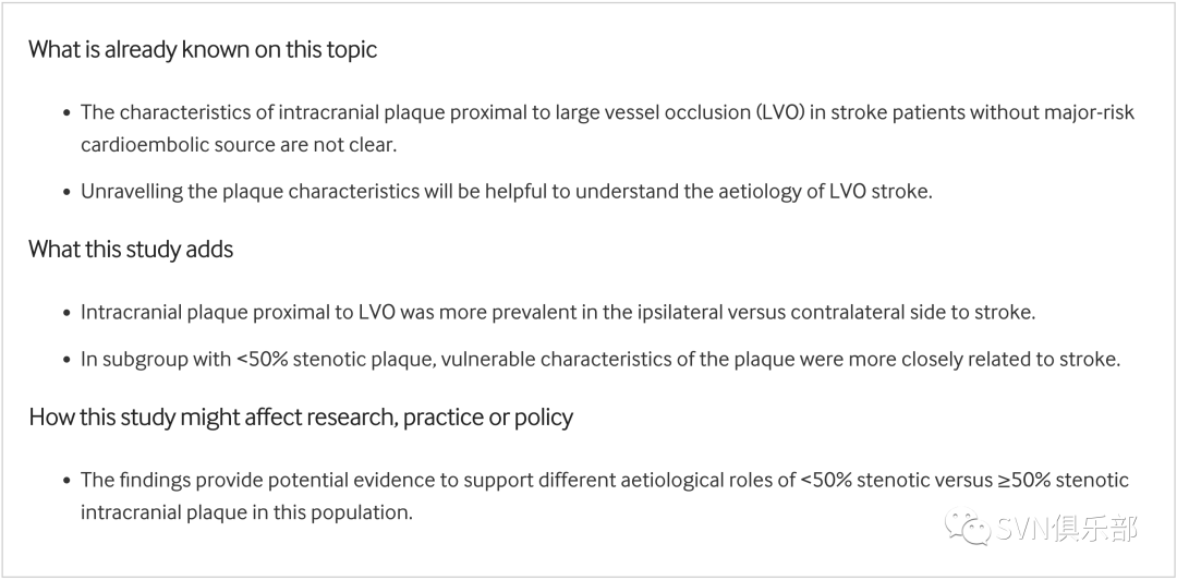
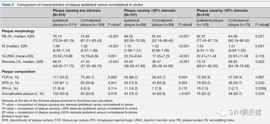
研究结果显示,在279例卒中患者中,LVO近端颅内斑块在卒中同侧相较于对侧更为普遍(75.6% vs 58.8%,p<0.001)。与卒中对侧相较,同侧斑块的PB(p<0.001)、RI(p<0.001)和%LRNC(p=0.001)较大,DPS(61.1% vs 50.6%,p=0.041)和复合斑块(63.0% vs 50.6%,p=0.016)发生率较高。Logistic回归分析显示,RI和PB与缺血性卒中的发生呈正相关(RI:粗比值比(crude OR)1.303,95%CI 1.072-1.584,p=0.008;PB:粗比值比(crude OR):1.677,95%CI 1.381-2.037,p<0.001)。在狭窄<50%的亚组中,较大的PB、RI、%LRNC和复合斑块的存在与卒中相关性更为密切,而在狭窄≥50%的亚组中则不明显。
本研究首次报道了非心源性卒中患者LVO近端颅内斑块的特征。这为支持该人群中狭窄<50%和狭窄≥50%的颅内斑块的不同病因学作用提供了潜在证据。
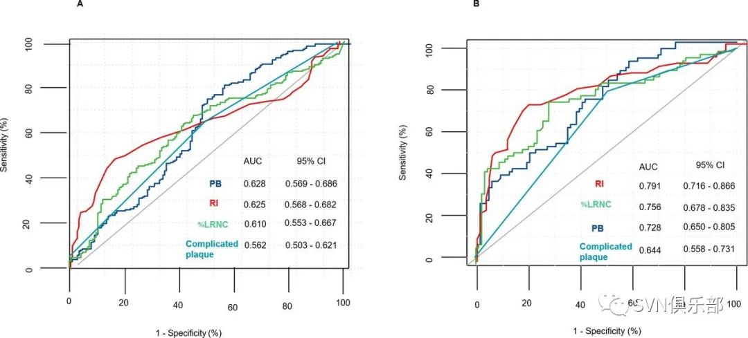
研究团队通过对受试者工作特征曲线(receiver operating characteristic curve, ROC)进行分析发现,在排除狭窄≥50%的斑块后,斑块生物标志物对缺血性卒中的鉴别能力显著改善。其中以RI的AUC(area under curve, AUC)增加最为明显(AUC(95%CI)从62.5%(56.8%~68.2%)增加至79.1%(71.6%~86.6%),两者间绝对差异区域:16.6%)(Figure 3)。
Figure 3. Receiver operating characteristics (ROC) analysis before and after excluding plaques with ≥50% lumen stenosis showing the changes of diagnostic performances of plaque biomarkers for predicting an index ischaemic stroke, before (A) and after (B) excluding plaques causing at least 50% lumen stenosis.
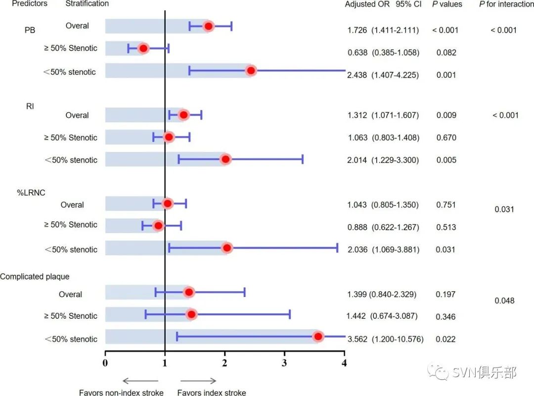
Figure 4. Subgroup analysis stratified by stenosis at the threshold of 50% caused by plaque showing the distinct characteristics of plaque ipsilateral vs contralateral to stroke in subgroup with ≥50% stenosis or <50% stenosis. The circles (red) and shadows (light blue) represented the points estimation and strengths of OR, respectively. The error bars (blue lines) represented DPS, discontinuity of plaque surface, LRNC, lipid rich necrotic core, PB, plaque burden, RI, remodelling index.
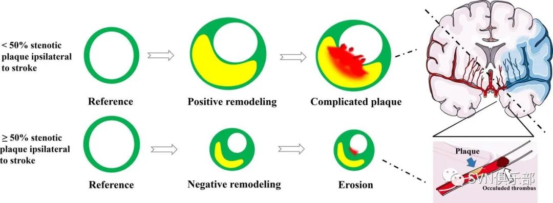
该研究发现,LVO近端同侧斑块的形态学特征(如PB和RI)是颅内LVO非心源性卒中人群中首发缺血性卒中的独立预测因子,这与之前的研究一致。研究发现RI和PB与卒中密切相关。同时还发现狭窄与斑块形态特征(如PB和RI)相互作用。这些结果表明,卒中事件的发生与高危斑块之间的相关性在斑块形成的不同阶段可能是动态的,表明急性LVO潜在的不同机制(Figure 5)。
Figure 5.The possible underlying aetiology of LVO in <50% stenosis versus ≥50% stenosis group In <50% stenosis group, a positive remodelling plaque was highly associated with increased vulnerability (disrupted fibrous cap, IPH or mural thrombus) and the index stroke, while in ≥50% stenosis group, a negative remodelling plaque with thick fibrous cap erosion to index stroke. Upper: showed a positive remodelling plaque with IPH and thin, ruptured fibrous cap ipsilateral to stroke. Lower: showed a negative remodelling plaque with thick fibrous cap erosion ipsilateral to stroke. Green=vessel wall/fibrous tissue, yellow=LRNC, red=IPH/mural thrombus.

来源:SVN俱乐部
转载已获授权,其他账号转载请联系原账号

专家共识 | 急性缺血性卒中替奈普酶静脉溶栓治疗中国专家共识
查看更多
 中国医学论坛报
 壹生
壹生 今日肿瘤
今日肿瘤 今日循环
今日循环 今日糖尿病
今日糖尿病 今日口腔
今日口腔 全科周刊
全科周刊 脱贫地区农副产品网络销售平台
京公网安备 11010202008182号
| 互联网新闻信息服务许可证编号:10120190017